
Burning or shooting pain describes what you feel, not why it is happening. By the end, you will understand how the pain map, neurological examination, medical history, laboratory tests, electrodiagnostic studies, and imaging are combined to distinguish nerve pain from problems in the spine, muscles, joints, skin, or blood vessels.
Key takeaways
- Pain location and travel can distinguish a nerve, nerve root, or wider disorder.
- Weakness, sudden numbness, or bladder changes require urgent neurological assessment.
- A normal scan or nerve test does not always rule out nerve pain.
- Neurologists combine symptoms, examination findings, illnesses, and test results before planning care.
How the pain pattern points to a nerve, nerve root, or wider disorder
Burning pain and shooting pain are descriptions, not diagnoses. A neurologist uses a nerve pain history to map where symptoms begin, where they travel, and whether the pain follows a single nerve, a nerve root, several peripheral nerves, or a wider brain or spinal-cord pattern.
The history records:
- Exact location and pain distribution, including whether symptoms stay in one strip, spread down an arm or leg, or affect both feet or hands
- One-sided versus two-sided symptoms
- Sudden or gradual onset, duration of each episode, and progression over days, weeks, or months
- Triggers such as walking, coughing, bending, movement, cold, or light touch
- Night-time worsening, pain from clothing or bedsheets, and relief from changing position
- Numbness, altered temperature sensation, weakness, imbalance, and associated back or neck pain
Pain that travels from the back into one leg with coughing or bending suggests irritation of a nerve root. Burning in both feet that slowly spreads upward points more toward peripheral neuropathy, while pain from light touch supports altered sensory processing but does not identify one disease.
A band of altered sensation, symptoms beyond one limb, bowel or bladder changes, or weakness on one side raises concern for spinal-cord or brain involvement. The pattern also helps separate nerve pain from joint, muscle, skin, or blood-vessel problems.
What the neurological examination can reveal
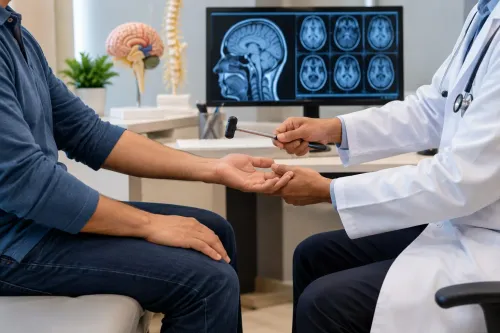
The neurological examination shows which nerve fibers and pathways are involved by comparing sensation, strength, reflexes, and movement on both sides. Pinprick and temperature mainly test small sensory fibers; vibration sense and joint-position testing assess large sensory fibers. Weakness or muscle wasting points to motor involvement.
| Finding | What it suggests | Important limit |
|---|---|---|
| Reduced pinprick or temperature | Small-fiber or peripheral nerve involvement | The pattern matters more than one spot |
| Loss of vibration sense | Large-fiber neuropathy or posterior-column disease | It can be normal in isolated small-fiber pain |
| Weakness or muscle wasting | Motor nerve, nerve root, or spinal-cord involvement | Pain alone does not cause true weakness |
| Absent ankle reflexes | Peripheral nerve or nerve-root disease | This finding alone does not identify the cause |
| Brisk reflexes or an abnormal plantar response | Spinal-cord or brain pathway involvement | It must match other examination findings |
The examiner also maps the border of numbness, checks gait and balance, and tests whether weakness follows one nerve, one root, or several nerves. A normal examination does not rule out neuropathic pain: small-fiber disorders can preserve strength, vibration sense, and reflexes, and symptoms can precede measurable deficits.
Repeated examination and targeted tests may therefore be necessary.
Which warning signs require urgent assessment
Burning or shooting pain needs urgent assessment when it arrives with a new neurological deficit, rapid progression, or loss of blood flow—not merely because the pain is severe.
| Warning sign | What it may indicate | What to do |
|---|---|---|
| Sudden weakness, facial droop, speech trouble, or one-sided numbness | Stroke or another acute brain problem | Call emergency services immediately |
| Bladder or bowel dysfunction, saddle-area numbness, or severe back pain with leg weakness | Spinal-cord or cauda equina compression | Seek emergency assessment now |
| Rapidly ascending numbness, worsening weakness, or trouble breathing | A rapidly progressive inflammatory or infectious neuropathy | Go to an emergency department |
| A cold, pale, blue, or pulseless painful limb | Blocked arterial blood flow | Seek emergency vascular care |
| Severe pain with fever, confusion, or marked illness | Infection, inflammation, or another serious systemic cause | Obtain urgent medical evaluation |
| A new blistering rash with burning pain, especially near an eye | Shingles affecting an important nerve | Arrange same-day medical care |
Do not wait for a routine consultation if symptoms are progressing over hours or days. A normal earlier scan or nerve test does not overrule a new deficit.
How other illnesses and pain conditions enter the investigation
Diabetes is common, but it is not the only metabolic explanation for burning feet or electric-shock pain. The neurologist matches each possibility to its timing, distribution, examination findings, and blood results.
- Vitamin B12 deficiency, especially with balance trouble or loss of vibration sense
- Thyroid disease, which can affect nerves and muscles
- Kidney disease, when waste products accumulate and irritate nerves
- Alcohol exposure or impaired glucose tolerance
- Chemotherapy neuropathy or another toxic medication effect
- Paraproteinemia, autoimmune disease, infection, or an inherited neuropathy
Burning sensations can also come from the brain or spinal cord rather than peripheral nerves. Multiple sclerosis, a spinal-cord lesion, or stroke becomes more concerning when symptoms cross several nerve territories, affect one side, create a sensory level, or accompany stiffness, abnormal reflexes, or bladder changes.
The clinician also tests whether the pain is nerve pain at all. Joint or tendon disease, muscle pain, vascular disease, and skin disorders can produce pain that feels deep, hot, or sharp. Allodynia and hyperalgesia show altered pain processing but do not identify one specific disease.
A rapidly worsening pattern with weakness or systemic illness calls for expedited assessment.
Which tests clarify the cause—and what a normal result means
A normal result narrows the possibilities; it does not prove that the pain is imaginary or unrelated to nerves. Each test examines a different structure, so the neurologist matches the result with your pain map, strength, reflexes, and sensory findings.
| Test | What it helps determine | What a normal result means |
|---|---|---|
| Blood tests | HbA1c checks for diabetes or poor glucose control; vitamin B12 testing looks for deficiency; serum protein electrophoresis screens for abnormal proteins linked to some neuropathies | The tested causes become less likely, but autoimmune, medication-related, hereditary, and small-fiber causes can remain |
| Nerve conduction study | Measures how quickly and strongly electrical signals travel through large peripheral nerves | Large-fiber neuropathy, nerve entrapment, or some nerve injuries become less likely; isolated small-fiber neuropathy can still cause genuine burning pain |
| Electromyography EMG | Examines muscle electrical activity for motor-unit damage and supports a diagnosis of radiculopathy or nerve injury | A major motor-root or muscle abnormality is less likely, but a recent or purely sensory problem can escape detection |
| Skin biopsy | Measures intraepidermal nerve-fiber density when small-fiber neuropathy is suspected | Reduced density supports small-fiber neuropathy; a normal sample does not identify or completely exclude every cause |
| MRI | Shows discs, spinal nerve roots, spinal cord, joints, masses, or other structural problems | No visible structural explanation appears; MRI does not measure small nerve fibers or prove that an abnormal disc causes pain |
A normal nerve conduction study and EMG therefore do not end the investigation. If burning pain and altered temperature or pinprick sensation persist, the clinician may consider skin biopsy or autonomic testing such as quantitative sudomotor axon reflex testing; these support small-fiber neuropathy but do not reveal its underlying cause.
How the neurologist combines conflicting findings into a plan
A likely cause is assigned by the pattern that remains consistent across the neurologist evaluation, not by the most striking test result. A normal examination or EMG does not erase convincing symptoms, especially when small-fiber neuropathy is suspected.
1. Start with the timeline. Pain that began yesterday after a back injury is weighed differently from burning in both feet that progressed over 18 months. Early testing can miss evolving nerve injury, so repeat examination or delayed EMG may be more informative.
2. Check whether findings agree anatomically. A radiculopathy diagnosis becomes stronger when limb pain follows one nerve-root territory and matches myotomal weakness, reduced reflexes, or sensory loss. Pain radiating from the spine alone is not enough.
3. Interpret tests as supporting evidence. Normal nerve-conduction studies can occur with small-fiber neuropathy; skin-biopsy nerve-fiber density or autonomic testing can support it, but neither identifies the underlying cause. Blood glucose, vitamin B12 with metabolites when appropriate, and serum protein immunofixation help screen for treatable contributors.
4. Treat imaging as a question, not an answer. An MRI disc bulge matters when it matches the side, level, examination, and timing of symptoms. Because disc bulges are common without pain, an unmatched scan should not redirect the whole plan.
Dr Jharna P Mahajan (Bhanushali) uses this kind of cross-checking to separate a test abnormality from the cause that best explains the patient’s pattern.
Frequently asked questions
How can pain patterns show whether pain comes from a nerve or nerve root?
A neurologist maps where pain starts and travels, then checks whether it follows one nerve, a nerve root, several peripheral nerves, or a wider brain or spinal-cord pattern.
What can a neurological examination reveal about burning or shooting pain?
Testing strength, reflexes, sensation, coordination, walking, and nerve distribution can reveal weakness, sensory loss, reflex changes, or signs of nerve-root involvement.
Which warning signs require urgent assessment for nerve pain?
Seek urgent assessment for sudden weakness or numbness, new trouble walking, loss of bladder or bowel control, numbness around the groin, or symptoms with severe back pain.
What does a normal nerve test or scan mean?
A normal result can reduce the likelihood of some nerve, root, or structural disorders without ruling out every cause of nerve pain; the result must be interpreted alongside symptoms and examination findings.
How are other illnesses and pain conditions included in the investigation?
The neurologist reviews conditions such as diabetes, thyroid disease, vitamin deficiencies, infections, medication effects, joint problems, and pain syndromes that can mimic or worsen nerve pain.